

Next-Gen Navigation: How Virtual Planning and 3D Prints Guide Surgeons to Surgical Success
It’s been almost 10 years since Ivy Chacon became the first patient at Cook Children’s Medical Center to benefit from the use of 3D technology to plan her rare and complicated cardiovascular surgery, and the surgical repairs made then, still hold today.
“There is no doubt the 3D print of Ivy’s heart that surgeons used to plan and prepare for her procedure contributed to the positive outcome and long-term success of the surgical repair,” said Steve Muyskens, M.D., cardiologist and medical director of cardiac MRI and the 3D Lab at Cook Children’s Medical Center–Fort Worth. “Three-dimensional printing takes away the guesswork for physicians, allowing them to completely visualize a patient’s anatomy and unique anomalies and map their intervention before they get to the operating room.”
Ivy, now 10, was born with congenitally corrected transposition of the great arteries (ccTGA) in which the two lower pumping chambers, or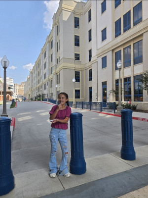 ventricles, of her heart are reversed. In a normal heart, the right atrium sends unoxygenated blood through the right ventricle, which pumps it through the pulmonary artery into the lungs. The blood is then oxygenated and flows back into the left atrium. From there, the left ventricle pumps the blood into the aorta where it is carried throughout the body.
In Ivy’s heart, however, the ventricles were switched. This meant the normally weaker and thinner-walled right ventricle was doing the high-pressure job of pumping blood throughout the body via the aorta—a function best suited for the thicker, stronger left ventricle. This inefficient pumping function put her at high risk for heart failure, irregular heart rhythms and sudden cardiac death. Ivy’s heart was further complicated by having a hole in the bottom chamber and a very small pulmonary valve, causing limited blood flow to her lungs. Ivy’s heart defects were unable to be repaired in infancy, so she had a palliative procedure at 2 weeks old to stabilize her condition while she grew.
Then, at 19 months old, Ivy became a candidate for the Nikaidoh procedure, also known as an aortic translocation. The procedure is named after Hisashi Nikaidoh, M.D., a retired Cook Children’s cardiovascular surgeon and innovator. Dr. Nikaidoh developed the complex open-heart surgery that reverses the translocation and reconstructs a more normal ventricular and arterial layout. He assisted Vincent Tam, M.D., during the surgery. Dr. Tam is a pediatric cardiothoracic surgeon and Cook Children’s medical director of Cardiothoracic Surgery.
To prepare for the procedure and to explain the complex surgery to Ivy’s family, Dr. Tam used a 3D printing of Ivy’s heart that was created in Cook Children’s 3D Lab. The printed heart allowed Dr. Tam to visualize Ivy’s exact anatomy so that he knew what to expect going into surgery.
It also helped Ivy’s family understand her complicated diagnosis and Dr. Tam’s plan to save her life.
“I've noticed over time that having these 3D models helps parents and other care providers, like nurses, better connect with the plan of care,” Dr. Muyskens said. “If you think about it from a parent standpoint, you're trusting this person, a surgeon or even just a cardiologist like myself, to take your child on a journey that's going to make them as happy and healthy as possible, oftentimes with no understanding of this very complex issue. So if you're able to then bridge that gap and have the surgeon show a parent an actual 3D model they can hold in their hands and say, ‘Here's your child's heart. This is why it's so complex. This is why it's different. There’s not a template for this, but this is what we think we can do. Let's walk through this together and figure out how we want to care for your child as a medical team and a family.’ I think that really helps connect those two pieces when you have a really unusual case.”
It certainly did for Ivy’s mom, Elizabeth. She says the physical model of Ivy’s heart helped her understand her baby’s complicated condition and Dr. Tam’s surgical plan. Having that knowledge was empowering and compelled her to hold on to hope during her daughter’s day-long surgery.
Surgical Solutions
In addition to his work in the 3D Lab, Dr. Muyskens is a cardiologist with a full patient load. He often partners with cardiothoracic surgeons to repair congenital heart defects and cardiovascular anomalies in his patients. When 2D imaging was the only resource for visualizing and understanding a patient’s anatomy, it wasn’t uncommon for Dr. Muyskens to hear surgeons say, “We’ll have to see for sure when we get in there,” when discussing a surgical intervention. This element of the unknown sparked a vision to create the 3D Lab to give physicians a deeper, more detailed look into the problems they were working to solve.
“I always felt like we need to be doing more and wondered how I can further the offering we have to help surgeons prepare for complex surgeries,” he said. “Your heart is about the size of your fist. So if you think about the size of a baby’s heart, and then trying to do open-heart surgery and move coronaries and great vessels and create new pathways for blood flow, it's a very complex surgery. By 3D printing the heart, we can have a much better understanding of those relationships prior to surgery. That's how we got started. Now we have three printers, surface scanners and virtual and augmented reality options. We've expanded in a lot of ways.’
While the 3D Lab originated with cardiology, its tools and resources are now available to multiple specialties. In the 10 years since the Lab’s debut with Ivy’s surgery, specialties such as plastic surgery, craniofacial surgery and orthopedics now utilize its capabilities to create precise, unique copies of spines, facial features, bone structures and more. These models can be printed in multiple colors and materials that are engineered to be more tissue-like, whether that’s bone or soft tissues. This creates a more realistic feel for a surgeon who wants to practice drilling into bone or cutting into tissue prior to a surgery.
Practice Makes Perfect
The 3D Lab has expanded its pre-operative practice options, too.
“We're still doing the printing that we have done, but we've also moved a lot more into virtual surgical and interventional planning,” Dr. Muyskens said. “For complicated cases, we want to have a pre-operative or interventional plan in place so we know, for example in the catheterization lab, the exact camera angles we need and what size stents we should use. Now we can virtually place stents during simulation and review our work. Sometimes we go down the pathway virtually and we realize that it's probably not a great case for the cath lab because it’s actually much trickier than the images would've suggested. So we will then send those patients to surgery. That's kind of been our current frontier.”
Much like a pilot in a flight simulator, virtual reality allows surgeons to see internal structures, plan their approach and practice their cuts in the Lab rather than saving these critical and time-consuming decisions for the OR when the patient is under anesthesia and the surgical site is open. This tangible experience outside of the OR builds valuable knowledge and spatial awareness of a patient’s unique anatomy, increases surgical accuracy, removes the element of surprise and decreases time under anesthesia—all precursors to improved outcomes for patients.
Mirror Image Models
The addition of a 4B printer allows the Lab to produce biocompatible materials that can be used temporarily in the sterile surgical field, like custom cutting guides or molds used to repair facial fractures.
Take a cheekbone fracture, for example. Classically, a surgeon would shape a piece of mesh to look similar to the other side, place the mesh inside the cheek area and attach the broken bones to the mesh so that they heal in approximately the same shape as the other side.
“Now we can take scans of the healthy side, mirror the image and print a 3D model,” Dr. Muyskens said. “Surgeons can then take that into the OR and prebend plates and mesh around the printed model to create the ideal version of the fractured side. Not only does it save time, but it allows the surgeon to create a repair that looks exactly like the healthy side.”
The 3D Lab’s use of surface scanners also contributes to improved aesthetic outcomes. By scanning an already 3D object, like an ear, the Lab can produce exact replicas that a surgeon can use to repair malformations.
Leveraging these tools to plan ahead and remove as much of the guesswork as possible leads to fewer complications and improved outcomes. It also translates to lower health care costs for patients and the health care system as a whole, according to Dr. Muyskens.
“We were pretty early adapters of this technology,” Dr. Muyskens said. “So I think it says a lot about Cook Children’s and our desire to provide innovative care and solve complex problems using state-of-the-art methods.”
10 Years and Counting
Today, Ivy is full of life. Thanks to good surgical planning made possible with the 3D Lab, expert execution by Dr. Tam and Dr. Nikaidoh, and ongoing care from Cook Children’s cardiologist Lisa Roten, M.D., Ivy’s heart is still going strong. She loves to skate, play with her dog and have fun.
“Her personality is so big,” Elizabeth said. “She acts like nothing happened to her. She acts like she's not even sick, and like she doesn't have a pacemaker. She does what she wants to do. She's like a little energizer bunny. Nonstop. She just keeps going.”
